Month: October 2019

Lab 8 – Plant DNA Extraction

This week, we extracted DNA from Erythranthe guttata leaf samples collected in the field (see Lab 3 for collection methods). The following methods are modified from Alexander et al. DNA extraction protocol.

  1. I labeled three 2.0 milliliter tubes with my sample codes: NPI-1, PRBW 006, and MONO 007.
  2. I added three sterile, 3.2-mm stainless steel beads to each tube.
  3. I added a small amount of dried leaf tissue from the field collection vials to each tube, making sure to clean the forceps between tubes to prevent contamination.
  4. My lab group loaded our samples into a modified reciprocating saw rack and mounted the rack to the saw. We turned on the saw, which shook the samples back d forth on speed 3 for 40 seconds.
  5. We briefly spun the tubes in a centrifuge for 20 seconds at full speed to pull the plant material down to the bottom of the tube.
  6. I added 434 microliters of the preheated grind buffer to each tube.
  7. Then, I incubated the buffered grandame at 65 degrees Celsius for 10 minutes in a hot water bath, mixing the tubes by inverting them every 3 minutes.
  8. Next, I added 130 microliters of 3M pH 4.7 potassium acetate, inverted the tubes several times, and incubated on ice for 5 minutes.
  9. I loaded the tubes in a centrifuge and spun at maximum speed (14,000) for 20 minutes.
  10. In the meantime, I labeled new 1.5mL micro centrifuge tubes with the sample IDs. I transferred the supernatant from the centrifuged samples into the new tubes. I avoided transfer of the precipitate.
  11. Then, I added 1.5x the sample volume of binding buffer (700 microliters) to each new sample tube.
  12. I added 650 microliters of these mixture to Epoch spin column tubes. I centrifuged these tubes for 10 min at 14,000rpm. When finished, I disposed of the flow-through in a hazardous waste container.
  13. I added 650 more microliters of the mixture from step 11 into the Epoch spin column tube, and repeated the spin and disposal of waste.
  14. To wash the DNA bound to the silica membrane, I added 500 microliters of 70% EtOH to the column and centrifuged at 14,000 rpm for 8 minutes. Then I disposed of the flow-through.
  15. I repeated the washing step, adding more EtOH, spinning, and discarding the waste.
  16. Then I ran another centrifuge spin with the tubes for 5 min at 14,000 rpm to dry out any residual ethanol.
  17. I placed the columns in new, sterile, labeled 1.5-mL micro centrifuge tubes.
  18. We added 100 microliters of preheated (65 degrees Celsius) pure sterile H2O to each tube.  We let them stand for 5 minutes, then centrifuged for 2 min at 14,000 rpm.
Lab 7 – Phylogenetic Inference

I finished the previous lab by conducting a BLAST search of the COI gene from 24 ray-finned fishes (Actinopterygii) and one shark (Chondrichthyes). I created alignment of these 25 COI genes, and my two samples EZ02 and EZ03.

I started this lab by cleaning up the alignment, and deleting any extra base pairs from each end. Next, I used the program jModelTest2 to find the best model for these sequences. I exported my alignment from Geneious in a relaxed Phyllip format (.phy), and loaded it into jModelTest2.

From this alignment, I computed the likelihood scores, keeping all defaults for this calculation. I then used two different methods to find the best model. I first tried the Akaike Information Criterion (AIC) method. The best resulting method from this analysis was TVM+G. Then I ran the Bayesian Information Criterion (BIC) method. The best model based on BIC was TPM2uf+G.

Next, I ran a Bayesian inference to create a tree using MrBayes in Geneious. I used ‘HKY85’ for the substitution model, and gamma for the rate variation. I selected the shark gene for the outgroup. I first used a chain length of 10,000, but later repeated the methods with a chain length of 3,000,000. I used an unconstrained branch length and shape parameter of 10.

Next, I used maximum likelihood to infer a phylogenetic tree of my aligned data set. I did this with the RAxML plugin for Geneious. I used the rapid bootstrap with rapid hill climbing method. I used the resulting file to create a consensus tree with a support threshold of 50%.

Then, I tried a different program for maximum likelihood, called PHYML.

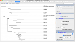
My final tree is shown in Figure 1.

Figure 1. Final Phylogeny

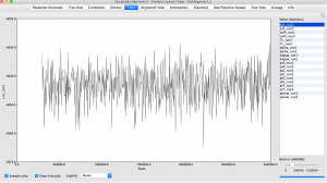
Figure 2. Phylogengy Trace

Figure 3. Parameter Estimates

Lab 6 – Intro to Geneious

This week, we worked with the program Geneious to sequence our fish DNA.  Unfortunately, the fish samples that I PCR’d did not produce a useable barcode sequence.  For this lab, I used barcodes from a previous student.  The samples were EZ02 and EZ03.  When she collected the sushi, the student was told that EZ02 was red snapper and EZ03 was yellowtail.  I used Geneious to assemble, edit and BLAST these DNA sequences, and found the results of the species barcodes.  EZ02 was not red snapper as advertised, it was Oreochromis niloticus, or Nile tilapia.  The other sample, EZ03 was Seriola quinqueradiata, or yellowtail, so this one was correctly labeled.


The EZ02 alignment I built had only one polymorphism, in column 663.  The EZ03 alignment had seven polymorphisms, found in columns 5, 6, 7, 11, 12, 15, and 201.

Viewing Message: 1 of 1.
Warning

Important: Read our blog and commenting guidelines before using the USF Blogs network.