Month: September 2019

Gel Electrophoresis and PCR Cleanup

In the second part of of the DNA extraction lab from animal tissue derived from sushi samples, our lab group ran a gel electrophoresis and PCR clean up. The first step in this process was obtaining our saved PCR tubes and letting them thaw at room temperature, and then the samples were placed on ice. In order to use our samples in gel electrophoresis, we pipetted 16 dots of dye on a sheet of parafilm. Each dot was approximately 1.5 microliters. We then pipetted 3 microliters of each PCR product into its own dot while changing pipette tips in between each dot. Each member was responsible for their own PCR products. After each dot of dye had PCR product in it, we set a pipette to  5 microliters and loaded each dot into our gel. We ran the gel at 130 volts for 30 minutes.

While our samples were running gel electrophoresis, we started our PCR cleanup. We began by carefully labeling new 0.2 microliter PCR tubes with our sample codes. I labeled my tubes with GS01, GS02, Gs03, and  GS04. We proceeded to make an ExoSap Master mix by mixing 211.8 microliters of pure water, 25 microliters of 10x buffer (Sap 10x), 88 microliters of SAP, and 4.4 microliters of Exo. We were able to do this successfully eventually, but initially, we did not use the correct amount of buffer or SAP, thus we had to redo our master mix. We mixed our successful master mix by holding the tube of master mix and waving our arm left and right on a horizontal plane, multiple times.

We pipetted 7.5 microliters of each PCR product into a clean, labeled, 0.2 microliter PCR tube. We also added 12.5 microliters of the master mix into the labeled PCR tube and then placed all PCR tubes into a thermocycler and started the EXOSAP program for approximately 45 minutes. When the program was completed, our professor placed the PCR tubes in a labeled rack and then they were placed in the freezer.

Field Trip on 9/10/19

Mimulus guttatus

On this field trip, our class traveled to Mountain Tamalpais where we were able to view the drastically different environments in which the species, Mimulus guttatus grows. We began the field trip by observing the shaded creek in which there was a large population of lively Mimulus guttatus. The environment consisted of a water bed from the creek and essentially no sunshine because of the shade created by all of the trees. This demonstrates how mimulus can grow without much sunlight. We were then able to collect samples of the species by pulling off the newest leaves from mimulus species that were at least 2 meters away from each other. The samples were places in a plastic tube with silica that would prevent moisture from accumulating and damaging the DNA sample.

This is the mimulus that I picked from, they were located in a shady creek

After collecting the samples, we traveled a short distance to a very dry area in the middle of a field where all plant life was very much exposed to the sun. In this area, there was a dried up creek that had some dried Mimulus guttatus that was previously seen alive. It is evident that the mimulus would have gotten the needed water from the creek that was there and was also equipped to handle large amounts of sunlight. We were also able to view the inside of a seed pod and view the seeds that species carries. They were incredibly small and thus it could be inferred that animal dispersal from feces or other matters were not a likely form of seed dispersal.

This is the area where the mimulus was dried because of sun exposure and a dried up creek.


These are the seeds from the mimulus guttatus, they are very small.

Next, we visited an area that plentiful with serpentine rock where plants have difficulty growing because of the heavy metals that come with the serpentine. The mimulus that were around this area were annual plants, meaning that the plants only grew once a year.

We then walked a further distance and reached an area in which there were many dry plants and sunlight exposure, but there was also a small creek that flowed in a valley. While the plants around the area were mostly dry, there was live mimulus guttatus where the stream was located. These species were flowering and the yellow color as well as the landing space of the petals suggested that bees were the pollinators that aided in seed dispersal of mimulus guttatus.

At the conclusion of the field trip, students were able to determine that Mimulus guttatus has been able to adapt to various environments. This can be due to gene flow that is transferred by bees and other pollinators that allows the plant to have certain traits that help the plant adapt.

DNA Extraction from Animal Tissue


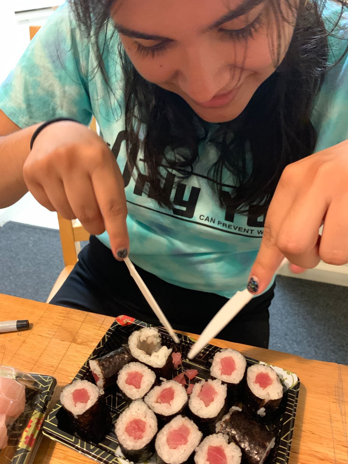
This picture shows the sushi at the Japanese market place

On September 2nd, 2019, I purchased packaged sushi from the Japanese grocery store, Nijya Market in San Francisco. On package was labeled to contain sushi that contained raw salmon and raw mackerel. Another package had raw tuna rolls, and the other had raw yellow tail. Pieces of the animal tissues from each fish were then placed into screw-cap microcentrifuge separately from each other and put on ice.

The image above shows myself removing the rice and other obvious ingredients from the raw tuna.

In order to extract DNA from fish tissue derived from sushi, it was first necessary to record the animal tissues that were collected on a data sheet. This included tuna, yellow tail, salmon, and mackerel. Each sample was given a unique code: tuna was labeled GS01, yellow tail was labeled GS02, salmon was labeled GS03, and mackerel was labeled GS04. Next, we put on gloves and labeled four 1.5 ml locking lid microcentrifuge tubes, one for each sample, with sharpies and writing on both the side and the top of the tube.

We cut a small piece of each sample using a scalpel and a paper plate as a surface. In order to determine the size of the sample piece that was cut, one of the samples was cut and weighed to 10 mg. The size of the 10 mg sample was then compared to other samples in order to create uniformity among the size of the samples.

Each sample was placed into its own 1.5 ml microcentrifuge tube. Before the addition of the sample piece, 100 microliters of Extraction Solution (ES) was added using a 200 micropipette and an unfiltered tip. 25 microliters of Tissue Preparation Solution (TPS) were also added using a 200 micropipette and unfiltered tips. The solution of ES and TPS was mixed by micropipetting up and down gently. The animal tissue sample was then placed in the corresponding labeled tube using forceps. The samples were mashed in each tube using a disposable non-filtered pipette tip. The samples in the tubes were then left alone at room temperature for 10 minutes.

The samples were placed on a heat block and incubated at 95 degrees Celsius for 3 minutes, using a phone as a timer. The sample was then removed from the heat block and and 100 microliters of Neutralizing Solution was added using a 200 micropipette with a filtered tip and mixed by vortexing. After this was completed, the samples were put on ice for approximately 4 minutes.

In order to assure good function of PCR, the gDNA was diluted by 10-fold. To do this, we labeled a microcentrifuge tube with “1:10” and the corresponding unique code on the side and top of the tube. We then added 18 microliters of purified water in the labeled tube along with 2 microliters of the gDNA that we extracted from the collected animal tissue. We also used the vortex machine to mix the solution.

We proceeded to make a master mix for the PCR that included 128 microliters of PCR quality water, 200 microliters of RED Extract, 16 microliters of forward primer, and 16 microliters of reverse primer. The volume was determined by multiplying the original volumes by 20 in order to accommodate for all members at our lab table and have extra for a negative control. We then used the master mix and placed 18 microliters of the master mix along with 2 microliters of our gDNA into a PCR tube. This process happened with each different gDNA while switching pipette tips in between each sample.

We wrote the labels of what gDNA it was on each PCR tube on the side and the top of the tube. The tubes then were placed in the thermocycler along with the negative control. Once the PCR reactions were all set up, the reaction ran for 1.5-2 hours. The PCR reactions were then placed in the freezer when the cycling was complete.

We are group Turtles, and the samples that are specifically mine preceded with “GS.” All samples were successful in producing a product.

Viewing Message: 1 of 1.
Warning

Important: Read our blog and commenting guidelines before using the USF Blogs network.