Month: November 2019

Lab 12 – Final PCR

Last week, we ran DD-RADSeq and Ligation on our Erythranthe guttata samples. This week, we began by running a test run of PCR to test for successful library construction.

  1. I began by labelling new PCR tubes to correspond to our samples #9-16 for this new PCR run.
  2. We created a Master Mix for the table, enough for 11 reactions, using the following recipe:
  • NEB One-Taq 2x Master Mix: 8uL/rxn (88 uL total)
  • Forward Primer (10mM): 0.4 uL/rxn (4.4 uL total)
  • Reverse Primer (10mM): 0.4 uL/rxn (4.4 uL total)
  • Pure H2O: 6.2uL/rxn (68.2 uL total)

3. We added 15 uL of the Master Mix and 1 uL of the library DNA template to each corresponding PCR tube.

4. We added these PCR tubes to the thermocycler BIORAD #1/2 –>DDRAD –>2_PCR1. This runs at 94 degrees Celsius for 2 min, then 20 cycles of (94 degrees C for 30 sec, 60 degrees C for 30 seconds, 68 degrees C for 45 sec). 4-10 degree C infinite hold.

Gel Run

  1. After PCR was finished, we ran the PCR products in a 1.5% agarose gel (0.75 g agarose in 50 mL 1XTAE) with a 100 bp ladder at 130V for 40 minutes.

Final PCR

  1. To add Illumina flow cell annealing sequences, multiplexing indices, and sequencing primer annealing regions to all fragments and to increase concentrations of sequencing libraries, we perform a PCR amplification with a Fusion Polymerase kit.
  2. We prepared a Master Mix 2 for the table, enough for 11 reactions, following this recipe:
  • Fusion DNA polymerase: 0.31 uL/rxn (3.4 uL total)
  • 5X Fusion HF Buffer: 6.25 uL/rxn (68.8 uL total)
  • 10uM Forward Primer (PCR1_X): 1.56 uL/rxn (17.2 uL total)
  • 10uM Reverse Primer (PCR2_5): 1.56 uL/rxn (17.2 uL total)
  • 10mM dNTPs: 0.63 uL/rxn (6.9 uL total)
  • DMSO: 0.94 uL/rxn (10.3 uL total)
  • Pure H2O: 10.8 uL/rxn (118.3 uL total)

4. For each reaction, we combined 3 uL of the library template DNA to a new, labeled PCR tube.

5. We then added 25 uL of the Master Mix 2 to each tube.

6. We vortexed, and then spun down in the centrifuge.

7. We ran PCR2 on thermocycler BIORAD #2. Specifically, this cycle ran as:

Cycle Step Cycles Temp Time
Initial Denaturation 1 98 degrees C 30 sec
Denaturation

Annealing

Extension

20 98 degrees C

65 degrees C

72 degrees C

10 sec

30 sec

30 sec (b/c 1kb genome)

Final Extension 1 72 degrees C 5 min
Hold 1 4 degrees C infinite


The next steps would be to run this PCR product on a gel and then select for bp size.
Lab 11 – DD-RADSeq

This week in lab we performed double-digest restriction associated DNA sequencing

Double Digest of Erythranthe guttate samples:

  1. To double digest 100-1000ng of high quality genomic DNA with selected restriction enzymes and appropriate digestion buffers, we performed the following steps.
  2. I labeled 8 PCR tubes 1-8, and added 6uL of each of the following samples’ DNA, to store on ice.
    1. MONO 005
    2. DIRA 007
    3. INVR 001
    4. CHIM 003
    5. CHIM 002
    6. PRBM 005
    7. CHIM 001
    8. PRBM 004
  3. We prepared a Master Mix for our table. Due to the small volumes used and the viscous nature of the glycerol the enzymes are stored in, we made 130% excess (enough for 11 reactions) of the following recipe:
  • 0.9 uL CutSmart 10X buffer/rxn   (9.9 uL total)
  • 0.28 uL EcoRI-HF enzyme/rxn.   (3.1 uL total)
  • 0.12 uL MPSI enzyme/rxn.           (1.32 uL total)
  • 1.7 uL pure H2O/rxn                     (18.7 uL total)

4. We stored the Master Mix 1 on ice.

5. We added 3 uL of Master Mix 1 to each sample’s DNA, changing pipette tips with every sample.

6. I sealed, vortexes and centrifuged the samples, and then added them to thermocycler with a heated lid set to 50 degrees Celsius, to incubate at 37 degrees Celsius for 8 hours (ran “DD” on BIORAD #1/2 –> DDRAD –>DD).


Adapter Ligation:

For this next step in the lab, we used samples that had previously undergone the first 6 steps, and were ready for adapter ligation.

  1. We pulled out the working stock EcoRI and Mspl adapters made previously, to thaw.
  2. We added 1 uL of the working stock EcoRI adapter directly to the digested DNA. We used different adapters in each sample, as paired below:

Sample 9 – Eco 2

Sample 10 – Eco 3

Sample 11 – Eco 4

Sample 12 – Eco 5

Sample 13 – Eco 6

Sample 14 – Eco 7

Sample 15 – Eco 8

Sample 16 – Eco 9

3. We prepared a Master Mix 2 for our table. Due to the small volumes used and the viscous nature of the glycerol the enzymes are stored in, we made 130% excess (enough for 11 reactions) of the following recipe:

  • 0.4 uL CutSmart 10X buffer/rxn                      (4.4 uL total)
  • 1.3 uL ATP 10mM/rxn                                        (14.3 uL total)
  • 0.2 uL T4 Ligase enzyme/rxn                            (2.2 uL total)
  • 0.7 uL pure H2O/rxn                                             (7.7 uL total)
  • 1.0 uL working stock universal P2 Mspl adapter  (11 uL)

4. I added 3 uL of Master Mix 2 to the digested DNA samples.

5. I sealed the samples, vortexes, centrifuged and added to a thermocycler with a heated lid set to 50 degrees Celsius to incubate at 16 degrees celsius for 6 hours (ran “LIGATE” of BIORAD #1/2 –>DDRAD –>LIGATE).

6. After the run, the samples were stored in a freezer.

Lab 10 – Erythranthe guttata PCR set up

In this week’s lab, we did a PCR run on our samples, using the following methods.

  1. We partnered up and labeled 6 PCR tubes P1-P6 (for Pyrimidudes 1-6), noting which label corresponds to which samples. Ours were labeled:

P1 – HWH CATB

P2 – MAPL 002

P3 – SHOR 001

P4 – NPI-1

P5 – MONO 007

P6 – PRBW 006

  1. We micro-pipetted 1 microliter of our samples of extracted DNA into the appropriate PCR tube.
  2. We created a master mix enough for 16 PCR reactions, for our lab group to share. This master mix consisted of:
    1. ddH2O (214 microliters)
    2. 10x buffer (32 microliters)
    3. MgCl2 (32 microliters)
    4. BSA (16 microliters)
    5. dNTPs (3.2 microliters)
    6. Forward primer (3.2 microliters)
    7. Reverse primer (3.2 microliters)
    8. Taq (0.64 microliters)
  3. We vortexed the master to mix it up. Then we added 19 microliters of the master mix to each of the PCR tubes with the DNA in them.
  4. These PCR tubes were then added to the thermocycler to run PCR.

Lab 9 – Mimulus DNA Gel

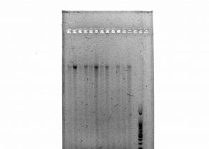
This week in lab, we ran our plant DNA that we extracted last week from Erythranthe guttata samples through a gel electrophoresis process.

We aloquoted 2-microliter drops of loading dye onto a piece of parafilm. Then, we added 3-4 microliters of our extracted DNA samples.  The entire solution was added to the gel cells, along with the other samples and a ladder in the final cell.  These samples were run in the gel for the remaining lecture (about an hour).

Figure 1. Photo of gel electrophoresis run by my lab group. My samples are the first three cells on the left.

Viewing Message: 1 of 1.
Warning

Important: Read our blog and commenting guidelines before using the USF Blogs network.