ISSR Scoring and Genomics Tutorial

During this lab we worked on reference based and de novo assembling. We examined the DNA sequences of various organisms and utilized tools found in Geneious to accurately assemble their DNA sequences. We also scored our ISSR analysis gels. These scorings will assist with comparisons later.


Reference Based Assembly questions:

  1. 4911, 4909, 4871, 497, 472,
  2. De Novo because paired-ends help researchers when there are no reference sequences. Although they can be used in reference sequences, the reference itself is a clue, whereas in de novo that clue is unavailable.
  3. 5.28 seconds
  4. average coverage: 98.1, max: 139. Lowest coverage came from the ends of the strands. This is probably because it’s harder to multiple fragments that line closer to the tips of the consensus sequence.
  5. 38,20,259,234. These sites are highly polymorphic
  6. 34->47, excluded because low coverage, invoking less confidence
  7. transitions: 4575,4567 transversions: 311, 4497
  8. OK
  9. 4499->4582 14 excluded.   293->310 none excluded
  10. 25,172 reads, 4 contigs, 17994,511,67096
  11. 41.7 seconds. Contig 4 low, contig 1 highest
  12. 90759->90774 & 92078->92266
  13. 24.4 seconds
  14. The consensus contained ambiguous bases when we would prefer mode bases. 58,851, 83786 & 268247
  15. 269124 bp.


ISSR Scores

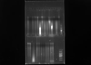
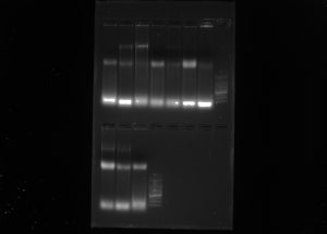
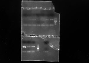
I referred to the short runs initially for bands and looked at the long runs to determine if there may have been any potential overlap. I also excluded individuals without both markers.

Pop. Gen Analysis I

Protocol:

  1. Added 1 microliter of loading dye to ISSR PCR products
  2. Loaded onto gel for imaging
  3. ran at 60 volts for about an hour and a half

Analysis of Lupinus ITS Sequences

  1. Downloaded Lupine_MEC_2017 file from Canvas
  2. Opened file in geneious
  3. Trimmed then assembled forward and reverse DNA sequences
  4. Selected all consensus sequences and made an alignment using default parameters of Muscle.

Results:

Write Up Questions

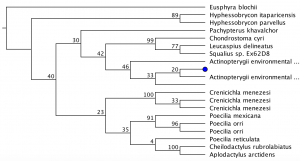
  1. Yes, my tree had one clade with a support value above .85. They were somewhat close together, and they all seemed to be from populations that had yellow flowers.
  2. Yes, a few populations that had purple flowers belonged to the same clade as other purple-flowered populations
  3. I believe that although ITS sequences can distinguish populations somewhat, the differences in these sequences are not enough to draw solid conclusions phylogenetically. This is because a lot of sequences failed to be grouped into distinct clades with other sequences. As a result of this, it is hard to claim that the ITS is useful enough to group these differing color populations phylogenically.

Plant DNA Analysis II

Protocol

  1. Added 1 microliter of loading dye to PCR products
  2. Ran PCR products in 1.3x agarose loading gel
  3. Ran for about 1 and a half hours at 60v
  4. Imaged gels to determine which ISSR’s work best
    1. Decided to use 17898 and OMAR
  5. Made 1:10 dilution of Plant DNA (10 microliters DNA, 90 Microliters water) from GWC samples
  6. Made two master mixes with
    1. ddh20        12.5 microliters
    2. 10x buffer + MH     3 microliters
    3. BSA            1 microliter
    4. dNTPs        2 microliters
    5. Primers     .25 microliters (either OMAR or 17898)
    6. tax              .25 microliters
    7. template    1 microliter
  7. Ran on thermocycler

ISSR Screening Test Run/psbA Amplification (Plant DNA Analysis I)

Inter simple sequence repeats (ISSR) uses microsatellite sequences as primers in PCR to generate multilocus markers that vary between organisms. Analysis and comparison of ISSR’s between organisms can reveal relatedness

Protocol:

Used ISSR markers (OMAR – (GAG)^4 RC) and psbA

On Samples (Left->Right)

GWC01 , CRA05 , PRO01 , PFR01 ,PRM01


20 tube Master mix for ISS (1 microliter template per tube)

ddH20 -> 250 microliters

10x buffer + Mg -> 60 microliters

BSA -> 20 microliters

dNTPs ->40 microliters

OMAR Primer -> 5 microliters

Taq -> 5 microliters


20 tube Master mix for psbA with following volumes in each tube

ddH20 -> 15.4 microliters

10x buffer + Mg -> 2 microliters

BSA -> 1 microliters

dNTPs ->.20 microliters

F Primer -> .2 microliters

R Primer -> .2 microliters

Taq -> .04 microliters

Placed on thermocycler


Plant DNA Extraction II

Protocol:

Used extracted DNA from the first Plant DNA extraction lab

GWC01-GWC05

  1. Loaded agarose gels with 3 microliters of template and 1 microliter of loading dye on parafilm
  2. ran for about 25 minutes at 130 v in the first bottom row of gel 2

Preparing template DNA:

  1. used two .2 ml pcr tubes
  2. pipetted 1 microliter of each sample into respective tube
  3. created master mix of (per tube) using primers of pSBA and ITS genes
    1. ddh20 – 15 microliters
    2. 10x buffer+mg – 2 μL
    3. BSA – 1 μL
    4. dNTPs – .20 μL
    5. f primer – .20 μL
    6. r primer – .20 μL
    7. Taq – .04 μL
    8. template – 1 μL
  4. Pipetted master mix into respective tubes and ran on thermocycler

Plant Tissue DNA Extraction

Processed

GWC01

GWC02

GWC03

GWC04

GWC05

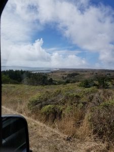
From Grey Whale Beach Site 3

Protocol:

  1. Labeled 1.5 ml centrifuge tubes with correct sample
  2. Added 3 sterile 3.2-mm stainless steel beads to each tube
  3. Added small amount of leaf tissue to each tube, wiped between
  4. Loaded tubes within a tube rack into the modified reciprocating saw rack and mounted rack into saw
  5. spun tubes down about 20 seconds on fast speed
  6. added 434 microliters of preheated grind buffer to each tube
  7. incubate buffered grandame at 65 degrees celsius for 10 min in water bath, mixing tubes by inversion every 3 mins
  8. add 130 microliters of 3M pH 4.7 potassium acetate, invert tubes several times and incubate on ice for 5 min
  9. Spin in a centrifuge at maximum force (about 14000-15000 rams) for 20 min
  10. Label new 1.5 ml tubes with sample ID
  11. Add 1.5 volumes binding buffer
  12. Add 650 microliters of mixture from step 11 to epoch spin column tubes and centrifuge for 10 min at 15,000 rpm in a centrifuge and discard flow-through
  13. repeat step 12 with the remaining volume from step 11
  14. wash with 500 ml of 70% EtOH then centrifuge at 15000 rpm until all liquid has passed through. discard flow-through
  15. repeat 14
  16. centrifuge columns at 15000 rpm for 5 more mins
  17. discard tube then place columns in 1.5 ml labeled micro centrifuge tube
  18. add 100 microliters preheated sterile h20 to each tube. let stand for 5 mins then centrifuge for 2 min at 15000 rpm to elute DNA

Gel Electrophoresis and Exo-Sap Clean Up


Protocol:

Gel Electrophoresis of gDNA

  1. Pipetted 2 microliters of loading dye per sample on parafilm (10 total)
  2. Added 3 microliters of PCR product to each loading dye dots
  3. Pipetted about 5 microliters into following wells
    1. RA01
    2. RA02
    3. RA03
    4. JPCA
    5. JPCB
    6. JPCC
    7. JPCD
    8. LADDER
    9. JW01
    10. JW02
    11. JW03
    12. LADDER
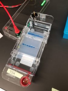
  4. Placed lid on pre-setup gel electrophoresis box with DNA running to red
  5. Set to 145 volts then ran for about 15 minutes

Imaged using gel doc EZ imager

Results: Not bad


Gel Electrophoresis of PCR Products

Making Gel:

  1. Small Gel- .5 g agarose, 50 ml 1x TAE Buffer, 0.5 microliters diluted Gel Red per 50 ml gel

However, we just reused the gel from earlier. It was melted in microwave for about 25 seconds. Then we added 1 microliter of Gel Red

2. Poured hot gel into gel mold, added combs and let cool


Gel Electrophoresis of PCR Amplified DNA

  1. Pipetted 2 microliters of loading dye per sample on parafilm (10 total)
  2. Added 3 microliters of PCR product to each loading dye dots
  3. Pipetted about 5 microliters into following wells
    1. RA01
    2. RA02
    3. RA03
    4. JPCA
    5. JPCB
    6. JPCC
    7. JPCD
    8. LADDER
    9. JW01
    10. JW02
    11. JW03
    12. LADDER
    13. NONE
    14. NEGATIVE
  4. Placed lid on pre-setup gel electrophoresis box with DNA running to red
  5. Set to 145 volts then ran for about 15 minutes

Imaged using gel doc EZ imager

Results:

Use RA02 RA03 JPCC JPCD JW01 JW02 JW03


ExoSap PCR Clean-Up Protocol