Lab 12: DD-RADSeq (PCR Test of Successful Library Construction)

Allyson Luber

November 19th, 2019

This lab session was combined with lecture for time efficiency. Order of class:

  1. PCR 1 TEST
  2. LECTURE
  3. GEL
  4. PCR 2  (final)

I. Test PCR: We tested for successful library construction of our Mimulus guttatus samples (restriction digest + ligation of barcodes) using a test PCR. PCR I: TESTS THE SUCCESS OF LIBRARY CONSTRUCTION. This PCR was performed with inexpensive non-high-fidelity Taq.

  1. Create Master Mix 1
  2. Ran PCR using PCR1 on BIORAD #1/2: 94 degrees celsius for 2 min, then 20 cycles of (94, 60, 68 degrees celsius for 30 seconds for all). 4-10 degrees celsius infinite hold
  3. After taken out of the PCR machine we ran the products of PCR 1 for each sample on a 1.5% agarose gel (o.75 g agarose in 50 mL TX TAE) with a 100 bp ladder at 130V for 40 minutes

Test PCR tubes (Samples 25-32)

Master Mix 1: Recipe for one PCR reaction of 16.0 μL. 10 rxn’s

  • NEB One-Taq 2x Master Mix (8.0  μL/1 rxn): 80 μL
  • Forward Primer (Primer 026, 10mM) (0.40 μL/1 rxn): 4.0  μL
  • Reverse Primer (10mM) (0.40 μL/1 rxn): 4.0 μL
  • Pure H2O (6.2 μL/1 rxn): 62  μL
  • Master Mix total: (15.00 μL/1 rxn) 150  μL
  • Library DNA Template (1.00 μL) –> total reaction volume 16.0 μL

II. Final PCR (PCR 2): This PCR run added the special “second barcode” sequences and the Illumina primers to our libraries of Mimulus gutattus, allowing us to identify which specific individuals a given sequence came from (Ligation barcode + PCR2 barcode). Each table used a different PCR2 primer. Our table: PCR2_7. PCR2: to generate final Illumina sequencing library

To add Illumina flowcell annealing sequences, multiplexing indices, and sequencing primer annealing regions to all fragments and to increase concentrations of sequencing, we performed a PCR amplification with a Phusion Polymerase kit

  1. For each library, we set up 4-8 PCR reactions (to combine and mitigate PCR bias) in 50 μL total volume
  2. For each PCR reaction, combine:
    • ~20 ng (~3  μL depending on concentration) of size-selected sample
    • PCR primers 1 and 2 at concentration 10 uM each
    • The recommended amount of 5X-HF buffer, 10 mM dNTPs, water, and Phusion DNAP
  3. Vortex, then spin down in microcentrifuge
  4. Run PCR2 on BIORAD #2 (Usually 10-20 cycles is sufficient). Increasing cycle number beyond this can introduce substantial mis-incorporation and exacerbate size and composition bias in final libraries
  5. Ran 2 μL of the products of PCR2 on a 1% agarose gel with a 100 bp ladder

Final PCR on samples 25-32 using PCR2_7

Master Mix 2 Recipe: One PCR rxn = 25.0  μL. 10 rxn’s made = ~220  μL

  • Phusion DNA Polymerase (o.31 μL/ 1 rxn): 3.1  μL
  • 5X Phusion HF buffer (6.25 μL/1 rxn): 63  μL
  • Forward primer (1.56 μL/ 1 rxn): 15.6  μL
  • Reverse primer (1.56  μL/ 1 rxn): 15.6  μL
  • DNTPs (0.63 μL/ 1 rxn): 6.3
  • DMSO (0.94 μL/ 1rxn) : 9.4  μL
  • Pure H2O (10.75  μL/ 1 rxn): 108  μL
  • Master Mix Total (22.0  μL/1 rxn): ~220  μL
  • Library DNA Template: 3.00  μL/1 rxn + MM Total = 25.0 μL/1 rxn
Lab 11: DD-RADSeq (Double-digest restriction associated DNA Sequencing)

Allyson Luber

November 12, 2019

DD-RADSeq (Double-digest restriction associated DNA Sequencing) Lab

I. Double Digest : digesting our M. guttatus samples. The objective of this part of the lab was to double digest 100-1000 ng of high quality genomic DNA with selected restriction enzymes, using a digestion buffer appropriate for both enzymes.

  1. Placed 6 μL of each of our sample’s DNA in a PCR tube, then stored it on ice
  2. Prepared Master Mix 1: Recipe for one double-digest reaction = 3.0 μL
  3. Added 3  μL of Master Mix 1 to each M. guttatus DNA sample
  4. We observed that the total reaction volume is now 9  μL.
  5. Next, we sealed the samples, vortexed, centrifuged, and incubated at 37 degrees Celsius for 8 hours on a thermocycler with a heated lid set to 50 degrees Celsius (run DD on BIORAD #11-2 –> DDRAD –> DD)

Master Mix 1 Recipe:

Master Mix: 11 RXN’s (due to the small volumes used and the viscous nature of the glycerol the enzymes are stored in, we made at least a 130% excess of MM1 to accommodate multiple rounds of pipetting)

  • CutSmart Buffer 10X (0.90  μL/1 rxn) : 9.9  μL
  • EcoRI-HF enzyme (0.28  μL/1 rxn): 3.08  μL
  • MSPI enzyme (0.12  μL/ 1 rxn): 1.32  μL
  • Pure H2O (1.70  μL/ 1 rxn): 18.7  μL
  • Master Mix total (3.00  μL/ 1 rxn) : 51.7  μL

Double Digest tubes:

  1. SAMPLE 7: MONO-002 (RICKY)
  2. SAMPLE 15: DIRA-010
  3. SAMPLE 24: LOTR-002 (ELI)
  4. SAMPLE 8: MONO-003
  5. SAMPLE 4: CHIM-004 (ALLYSON)
  6. SAMPLE 16: PRBN-001

I. Adapter Ligation: Performed all steps with samples in ice

  1. I added 1  μL of the working stock EcoRI adapter directly to the digested DNA
  2. Due to the small volumes used and the viscous nature of the glycerol the enzymes were stored in, we made at least a 130% excess of master mix 2 to accommodate multiple rounds of pipetting
  3. Prepared MM2
  4. Added 3  μL of MM2 to the digested DNA
  5. The total reaction volume is now 13 μL. (Dr. Paul did this part: incubate at 16 degrees celsius for 6 hours on a thermocycler with a heated lid set to 50 degrees celsius). After ligation the samples can be stored frozen for a few weeks, if needed.

Ligation tubes

  1. SAMPLE 25: ECO-2
  2. SAMPLE 26: ECO-3
  3. SAMPLE 27: ECO-4
  4. SAMPLE 28: ECO-5
  5. SAMPLE 29: ECO-6
  6. SAMPLE 30: ECO-7
  7. SAMPLE 31: ECO-8
  8. SAMPLE 32: ECO-9

Adapter Ligation Master Mix recipe for RADseq (Master Mix 2): 11rxn’s. Before making this Master Mix, we added 1  μL of the EcoRI adapter assigned to us to each of our digested DNA products. Recipe for one adapter ligation reaction is 3.0  μL

  • CutSmart buffer 10X (0.40 μL/1 rxn): 4.4  μL
  • ATP (10mM) (1.30 μL/1 rxn): 14.3  μL
  • T4 Ligase (0.20  μL/1 rxn): 2.2  μL
  • Pure H2O (0.10  μL/ 1rxn): 1.1
  • Universal P2 MspI adapter (1.00 μL/1  μL): 11  μL
  • Master Mix total (3.00  μL/1 rxn): 33  μL

Lab 10: PCR Reaction with M. guttatus

Allyson Luber

November 5, 2019

  • In this lab session we spent the majority of our time preparing the master mix to put into our small PCR tubes
  • The master mix ratio had to be 19.00  μL for each reaction, but since we are a table of 3, we multiplied each ingredient to 11 to produce enough Master mix for each person

Tubes/Specimen ID

  1.  DIRA-012
  2. CHIM-007
  3. CATB-RG
  4. RWCKAJC
  5. MAPLAJC
  6. CATB-EVR
  7. AEL-CATB
  8. MAPL-001
  9. RDRK-003

(Tubes 1-3 is Ricky’s; 4-6 is Eli’s; 7-9 is mine)

Master Mix: PCR Reactions (20  μL)

Ingredients / per rxn ( μL) / rxn x 11 ( μL)

  • ddH2O / 13.36 / 146.9
  • 10x buffer / 2.00 / 22
  • MgCl2 / 2.00 / 22
  • BSA / 1.00 / 11
  • dNTPs / 0.20 / 2.2
  • F-Primer (Miri 70) / 0.20 / 2.2
  • R-Primer (Miri 70) / 0.20 / 2.2
  • Taq / 0.04 / 0.41
  • Template / 1.00 / n/a

Total: 19.00 per rxn / 11 rxn’s (our group’s total): 208.91  μL

  • With the master mix finished, we pipeted 19.00  μL to each of our individual rxn’s


Lab 07: Phylogenetic Inference

Allyson Luber

October 8, 2019

Lab 07: Phylogenetic Inference

In this lab we continued our work using Geneious program on the computer. Today we worked on our COI sequences a little more, including usage of another program which I’ll introduce later.

  1. Clean up alignment of COI sequences that includes fish DNA barcode and sequences that was downloaded from Geneious last week. Alignment should look neat and almost uniformal.
  2. Next, we used a different program called jModelTest2 to figure out the best model of molecular evolution for our sequences. JModelTest2 was downloaded from a website provided by our professor.
  3. Back to Geneious, we had to export our alignment in Phylip format, then back at jModel, export the file.
  4. In jModel, we worked with our alignment for awhile. First we computed likelihood scores. When they were done we chose the best model based on some optimality criterion. The two methods were Akaike Information Criterion (AIC) and Bayesian Information Criterion (BIC).
  5. For both methods, the analysis was done instantaneously where we were then guided to look at the results table where all the scores for the various models were shown.

Bayesian Inference

  1. Back in Geneious we selected a MrBayes analysis for a tree builder. Before we could do this we changed a couple things such as substitution model, rate variation, outgroup, and MCMC settings. Changing it will depend on what your professor prefers.
  2. Ran the analysis — it was quick!
  3. This time, we ran the analysis longer using 1,100,000 for the “chain length” and 100,000 for the burn-in. After the analysis was finished, the posterior output should be different from the first

Maximum Likelihood

  1. This last part of lab required using maximum likelihood to infer a phylogenetic tree of our aligned data set. In Geneious, download RAxML under tools –> plug-ins
  2. Next, under evolutionary model we chose “Rapid bootstrap with rapid hill climbing”
  3. Once RAxML was finished a new line was created and from here we had to build a consensus tree using that new line.
  4. The same procedure was repeated but instead of using RAxML we used PHYML (downloaded it first under plug-ins)
  5. Used HKY85 model of molecular evolution for this our the final Bayesian tree

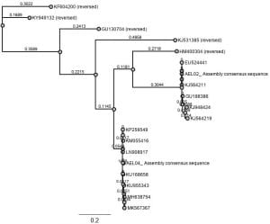
Final tree result

Lab 09: Gel Electrophoresis

Allyson Luber

10/29/2019

Lab 09:

We didn’t quite have a formal lab today, due to rescheduling purposes, but what we did instead was load our DNA samples from the previous weeks lab (Mimulus guttatus extractions) into the gel electrophoresis while we continued with lecture.

For each of our samples we loaded it with 2 μl of loading dye + 3 μl of our sample into each cell in the gel electrophoresis.

Order of lanes (L–>R)

  1. DIRA – 012
  2. CATD – RG
  3. CHIM – 007
  4. AEL – CATB
  5. MAPL – 001
  6. RDRK – 003
  7. EVR – CATB
  8. MAPL – AIC
  9. RWCK – AJC

#4-6 are mine.