Lab: Tuesday, 9/24/19
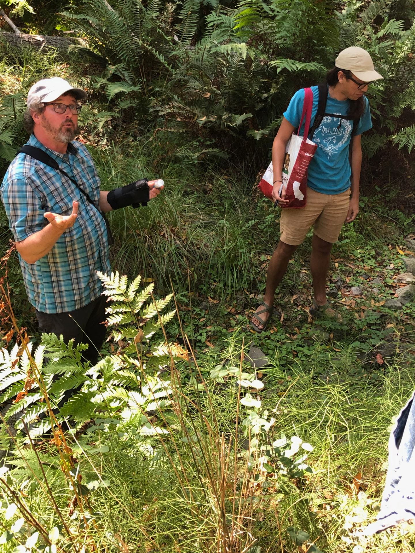
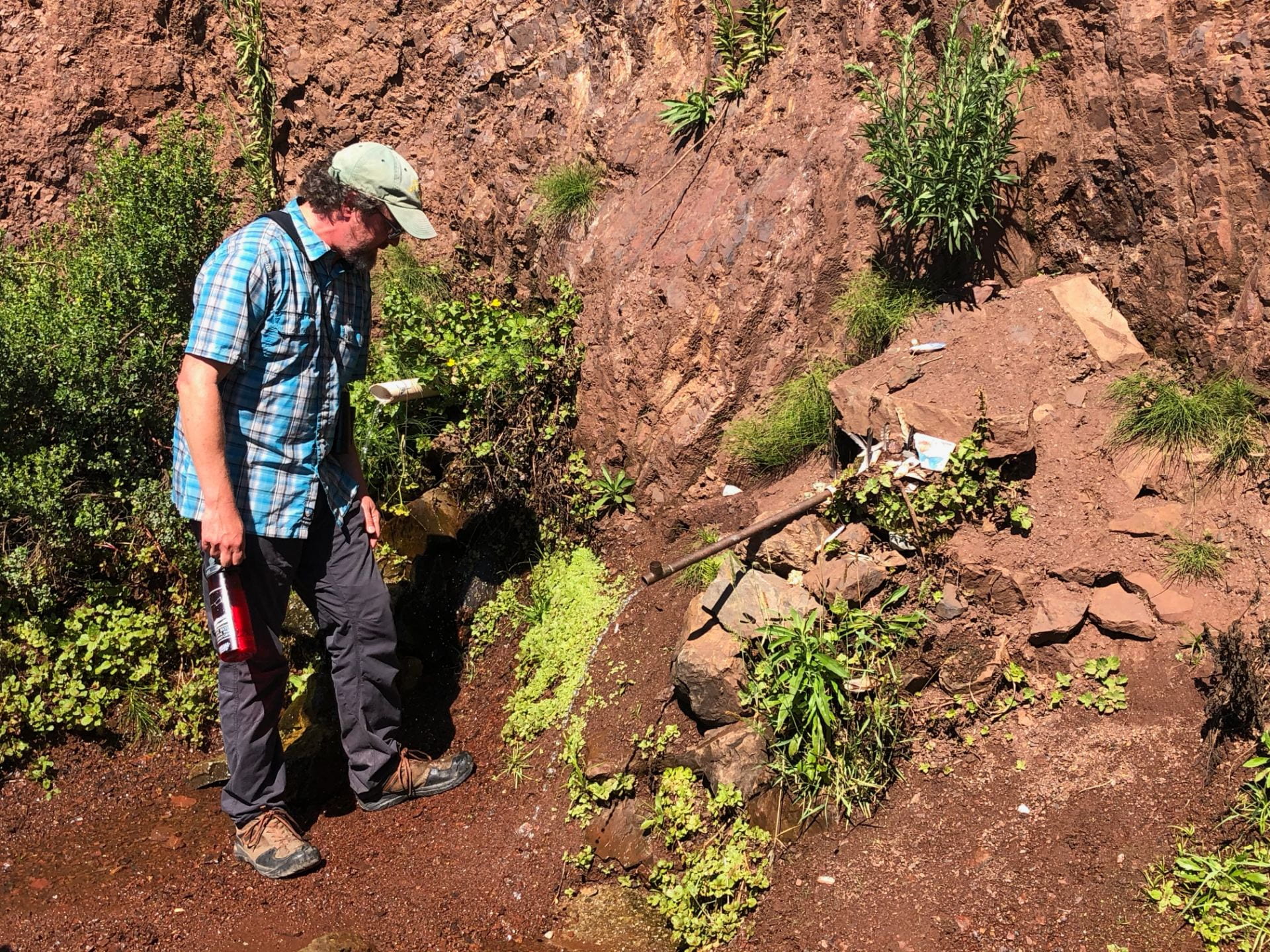
Location: Mt. Tamalpais
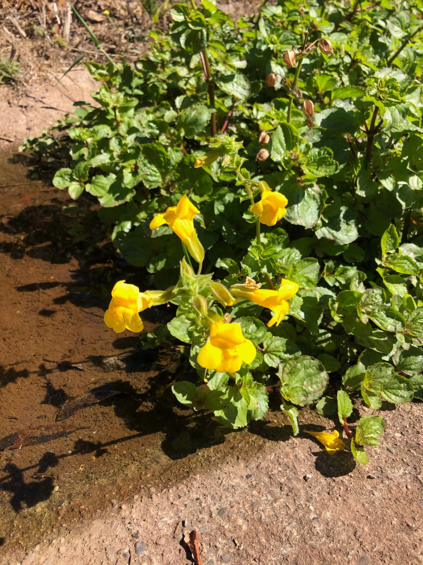
- Saw flowering population of mimulus that had really big flowers by the mountain water and stays moist throughout the summer.
Redwood Creek Mimuli
- Dynamic place
- Very low population density of mimulus
- Gene flow and seed flow were both primarily downstream
- Unlikely getting cues to flower with a very minimal interaction
- Conservation
DNA Extraction protocol:
- Labeled 3 2.0 mL with my sample codes: Tube #1 MM CATB, Tube #2 MM LOTR003, Tube #3 SCHO 003.
- Added 3 sterile 3.2 mm stainless steel beads to each tube.
- Added a small amount of leaf tissue to each tube – be sure to clean any tweezers or scalpels used between tubes to avoid cross-contamination.
- Loaded the tubes within a tube rack into the modified reciprocating saw rack and mount the rack to the saw.
- Spun down the tubes in the centrifuge (15-2o seconds at fast speed) to pull plant dust down from the lids.
- Added 434 µL preheated grind buffer to each tube.
- Incubated buffered grindate at 65C for 10 minutes in water bath, mixing the tubes by inversion every 3 minutes.
- Added 130 µL 3M pH 4.7 potassium acetate, inverted tubes several times and incubated on ice for 5 mins.
- Spun in the centrifuge at maximum force for 20 min. This will be 14,000 or 15,000 rpm for the tubes in a small microcentrifuge. Be sure to balance the microcentrifuge.
- Labeled new 1.5 mL tubes with the sample ID. Transferred supernatant to these sterile 1.5 mL microcentrifuge tubes. Avoided transfer of any precipitate. The precipitate will be fairly obvious and looked like small green particles on the bottom of the tubes or plate wells.
- Added 1.5 volumes binding buffer. Typically ~400 µL is recovered from previous steps, in which case 600 µL binding buffer is added.
- Applied 650 µL from Step 11 to Epoch spin column tubes and centrifuged for 10 minutes (until all liquid has passed through) at 15,000 rpm in a centrifuge and discarded flow-through.
- Disposed this buffer
13. Repeated step 12 with the remaining volume from step 11.
14. Washed the DNA bound to the silica membrane by adding 500 µL of 70% EtOH to the column and centrifuge at 15,000 rpm until all liquid has passed to the collection tube (8 mins). Discarded the flow-through.
15. Repeated step 14.
16. After discarding the flow-through from step 15, centrifuged the columns at 15,000 rpm for an additional 5 min to remove any residual ethanol.
17. Discarded the collection tubes and placed the columns in sterile 1.5 mL microcentrifuge tubes. *Made sure these 1.5 mL tubes are labeled with Sample ID and date — hold your final extracted DNA.
18. Added 100 µL preheated (65C) pure sterile H2O to each tube. Let stand 5 minutes and then centrifuged for 2 mins at 15,000 rpm to elute the DNA.
Tues, October 30th 2019
Gel Electrophoresis/PCR Cleanup
Gel Electrophoresis of PCR products:
- Grabbed PCR tubes from ice and thawed
- Dotted out 12 loading dye dots (2µL) on a sheet of parafilm
- Pipetted 3 µL of each PCR product into its own dot
- Loaded all the dots into the gel (7 µL each)
- Ran the gel at 130 volt for 30 minutes
Tues, November 5th 2019
PCR Reaction
Made up a Master Mix per table: *multiplied everything by 18
*Followed the protocol
Reagent Volume (µL)
ddH2O 240.48
10x Buffer 36
MgCl2 36
BSA 18
dNTPs 3.6
F-primer 3.6
R-primer 3.6
Taq 0.8
Template n/a
Total : 20 mL per rxn
Tubes Specimen ID
QS1 MM1
QS2 MM2
QS3 MM3
QS4 OY01
QS5 OY02
QS6 OY03
QS7 EB
QS8 RDRK001
QS9 SHOR005
QS10 KRS1
QS11 KRS2
QS12 KRS3
Tuesday, November 12th 2019
DD-RADSeq (Double-digest restriction associated DNA sequencing)
I. Double Digest
- Double digested 100-1000 ng of high quality genomic DNA with selected restriction enzymes. A digestion buffer was used for both enzymes.
- Placed 6 µL of each sample’s DNA in the PCR tube, stored on ice.
Sample #1: MONO 006 10
Sample #2: CHIM 005 5
3. Prepared Master Mix 1:
Added 3 µL of the Master Mix 1 to each sample’s DNA. (11 rxns)
CutSmart buffer 10x 0.90 µL x 11 = 9.9 µL
EcoRI-HF enzyme 0.28 µL x 11 = 3.08 µL
MSPI enzyme 0.12 µL x 11 = 1.32 µL
Pure water 1.70 µL x 11 = 18.7 µL
Master Mix total: 3 µL x 11 = 33 µL
4. Sealed the samples, vortexed, centrifuged, and incubated at 37C for 8 hours on a thermocycler with a heated lid set to 50C.
II. Adaptor Ligation
- Thawed the working stock EcoRI and MspI adaptors made previously.
Sample #
1 – 17: Eco-2
1- 18: Eco-2
1 – 19: Eco-3
1- 20: Eco-3
1 – 21: Eco-4
1- 22: Eco-4
1 – 23: Eco-5
1- 24: Eco-5
2. Added 1 µL of the working stock EcoRI adaptor directly to the digested DNA.
3. Prepared Master Mix 2
Added 3 µL of the Master Mix 2 to each sample’s digested DNA. (11 rxns)
CutSmart buffer 10x 0.40 µL x 11 = 4.4 µL
ATP (10 mM) 1.30 µL x 11 = 14.3 µL
T4 Ligase 0.20 µL x 11 = 2.2 µL
Pure water 0.10 µL x 11 = 1.1 µL
Universal P2 MspI adaptor 1.00 µL x 11 = 11 µL
Master Mix total: 3 µL x 11 = 33 µL
4. Sealed the samples, vortexed, centrifuged, and incubated at 16C for 6 hours on a thermocycler with a heated lid set to 50C.
Tuesday, November 18th 2019
DD-RadSeq (PCR Test of Successful Library Construction)
I. Test PCR
Tested for successful library construction of Mimulus gutattus samples (restriction digest and barcode ligation) using a test PCR.
Steps
PCR I will test for the success of library construction and can be performed with inexpensive non-high-fidelity Taq.
- Prepared a Master Mix for one PCR reaction of 16.0 µL
Master Mix: Rxn: 1 Rxns: 11
NEB One-Taq 2x Master Mix 8.0 µL 88 µL
Forward Primer (10 mM) 0.40 µL 4.4 µL
Reverse Primer (1o mM) 0.40 µL 4.4 µL
Pure H20 6.2 µL 68.2 µL
Master Mix Total 15.0 µL 165 µL
Library DNA Template 1.00 µL
Total rxn volume 16.0 µL
Test Samples: 17, 18, 19, 20, 21, 22, 23, 24
2. Ran the PCR using “PCR1” on BIORAD #1/2 (BIORAD #1/2 → DDRAD → 2_PCR1; the folowing conditions on a thermocycle with a heated lid: 94C for 30 sec, 60C for 30 sec, 68C for 45 sec). 4-10C infite hold.
3. Ran the products of PCR I for each sample on a 1.5% agarose gel (0.75 g agarose in 50 mL 1x TAE) with a 100 bp ladder at 130V for 40 minutes.
II. Final PCR (PCR 2)
This PCR run added the special ‘second barcode’ sequences and Illumina primer to Mimulus gutattus, allowing to identify which specific individuals in a given sequence comes from (Ligation barcode + PCR2 barcode). Each table used a different PCR2 primer. Our was PCR2_1. A PCR amplification was done with a Phusion Polymerase kit and added Illumina flowcell annealing sequences, multiplexing indices, and sequencing primer annealing regions to all fragments and to increase concentrations of sequencing libraries.
- For each library, 4-8 PCR reactions were set up in 50 µL total volume and prepared a Master Mix for one PCR reaction of 25.0 µL
Master Mix: Rxn: 1 Rxns: 11
Phusion DNA Polymerase 0.31 µL 3.41 µL
5X Phusion HF buffer 6.25 µL 69 µL
Forward Primer (1o mM) 1.56 µL 17.16 µL *PCR1_X)
Reverse Primer (10 mM) 1.56 µL 17.16 µL *PCR2_1
DNTPs (10 mM) 0.63 µL 6.93 µL
DMSO 0.94 µL 10.3 µL
Pure H2O 10.75 µL 118 µL
Master Mix Total 22.0 µL 242 µL
Library DNA Template 3.00 µL
Total rxn volume 25.0 µL
Test Samples: 17, 18, 19, 20, 21, 22, 23, 24
2. Vortexed and spinned down in microcentrifuge.
3. Ran “PCR2” on BIORAD #2 (BIORAD #2 → DDRAD → PCR2). Usually 10-20 cycle is sufficent. Increasing the cycle number beyond this can introduce substantial (>1%) base mis-incorporation and exacerabe the size and composition bias in the final libraries.
Cycle Step Cycles Temp Time
Initial denaturation 1 98C 30 sec
Denaturation 2 98C 10 sec
Annealing 65C 30 sec
Extension 72C 30 sec (b/c 1 kb genome)
Final extension 1 72C 5 minutes
Hold 1 4C
4. Ran 2 µL of the products of PCR 2 on a 1% agarose gel with a 100 bp ladder. Successful amplification should produce a faint smear of the fragment range that was targeted during size selection.